anti-MIN antibody information

INTRODUCTION

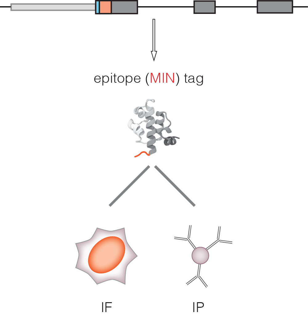
Once a MIN-tagged cell line is established, in-frame fusion of the MIN-tag to the target gene also results in the expression of a novel epitope tag. This epitope tag can be detected by a highly specific anti-MIN antibody, which can be used to screen for positive clones, perform co-immunoprecipitation experiments, as well as conventional and super resolution microscopy.
To request the MIN-tag antibody please visit the Request material page for further instructions
For more information see Mulholland et al. 2015

Detailed instructions and protocols can be found HERE

MIN_strategy

Using the MIN-antibody for Immunoprecipitation (IP)

Couple the MIN-antibody to beads

The monoclonal rat-anti-MIN antibody is provided as hybridoma culture supernatant. For coupling of the antibody to protein G beads (magnetic or sepharose), use a 10:1 ratio of antibody to bead volume, e.g. 500 μ antibody + 50 μ beads.

  • incubate beads with appropriate amount of antibody for 1 h at 4 °C while rotating
  • remove supernatant
  • wash twice with TBS-T (20 mM TrisHCl pH 7.5, 150 mM NaCl, 0.02 % Tween), 10:1 ratio TBS-T:beads
  • store antibody-coupled beads in TBS-T at 4 °C
  • Immunoprecipitation

    In principle, any standard cell lysis procedure should be compatible with anti-MIN-IP. Usually, 25 μl beads are sufficient to precipitate MIN-tagged protein from ~ 10 million ES cells.

  • Lyse ES cells on ice; e.g. 10 million cells in 100 μl lysis buffer (20 mM TrisHCl pH 7.5, 150 mM NaCl, 0.5 mM EDTA, 0.5 % NP-40, 2 mM PMSF, protease inhibitor mix) for 30 min
  • Dependent on lysis protocol and protein of interest: precipitate cellular debris by centrifugation: 15 min, 14 000 g, 4 °C
  • Dilute cell lysate to 400 μl with IP buffer (20 mM TrisHCl pH 7.5, 150 mM NaCl, 0.5 mM EDTA) and incubate with 25 μl anti-MIN-coupled beads for 1 h at 4 °C while rotating
  • Remove supernatant
  • Wash 3 times with 500 μl wash buffer (20 mM TrisHCl pH 7.5, 250 mM NaCl, 0.5 mM EDTA)
  • For subsequent Western blot analysis: boil beads in 50 μl 2xLaemmli sample buffer at 95 °C for 10 min
  • Using the MIN-tag antibody in Immunofluorescence (IF)

    Immunostaining Samples with MIN-tag antibody

    The monoclonal rat-anti-MIN antibody is provided as hybridoma culture supernatant. For coupling of the antibody to protein G beads (magnetic or sepharose), use a 10:1 ratio of antibody to bead volume, e.g. 500 μ antibody + 50 μ beads.

  • Seed cells on coverslips at least 24h prior to fixation
  • Wash cells 2x with PBS (tissue culture grade, free of sodium azide)
  • Fix cells in 2%-4% formaldehyde in PBS for 10 min at room temp.
  • Exchange fixative stepwise with PBST
  • Wash 2x with PBST
  • Quench with freshly prepared 20 mM glycine dissolved in PBS for 10-20 min to block free aldehydes
  • Wash 2x with PBST
  • Permeabilize with 0.5% Triton X-100 in PBS for 10 min
  • Block for 1 hr in blocking buffer (1x PBST, 2% BSA, 0.5% Fish skin gelatin)
  • Wash 3x with PBST
  • Incubate for 5-10 min in blocking buffer while preparing antibody solutions
  • Incubate coverslips with primary antibody for 1 h in humidified chamber
  • Use the MIN-tag antibody in a 1:1000 dilution
  • Wash 3x with PBST
  • Incubate coverslips with appropriately diluted primary antibody for 1 h in humidified chamber
  • Wash 3x with PBST
  • Wash 2x with PBS
  • Postfix with 4% Formaldehyde in PBS for 10 min at room temp.
  • Exchange fixative stepwise with PBST
  • Counterstain DNA with DAPI (2 μg/ml in PBST) for 6 min at room temp.
  • Wash 3x with PBS
  • Rinse briefly with ddH2O, only immediately prior to mounting
  • Remove excess liquid by touching the coverslip edge to a Kimwipe tissue
  • Mount the coverslip on a drop of Vectashield